ABSTRACT
As an innovative as well as an interdisciplinary research project, this study performed an analysis of brain signals so as to establish BrainIC as an auxiliary tool for physician diagnosis. Cognition behavior sciences, embedded technology, system on chips (SOC) design and physiological signal processing are integrated in this work. Moreover, a chip is built for real-time electroencephalography (EEG) processing purposes and a Brain Electrical Activity Mapping (BEAM) system, and a knowledge database is constructed to diagnose psychosis and body challenges in learning various behaviors and signals antithesis by a fuzzy inference engine. This work is completed with a medical support system developed for the mentally disabled or the elderly abled.
LITERATURES SURVEY
The purpose of this work is to develop a physical EEG signal acquisition SOC for BEAM analysis. There are a growing number of studies on this issue, among which a diagnostic criterion on human health is suggested based on physiological feature extractions and facial expression recognition, and a forecasting technique thereof is proposed which is applied to a medical monitoring system.
SYSTEM FRAMEWORK
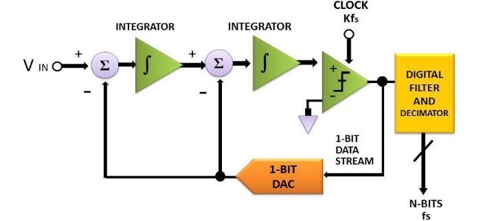
Figure 5. The schematic block diagram of a second order Σ-Δ AD converter with fs representing the sampling rate
A clear advantage gained is that high efficient analog and digital signal processing’s can be integrated with ease for the reason that most of the conversion is performed in the digital domain. Involving a comparator, an integrator and a 1-bit, dual output digital to analog converter, a Σ-Δ converter is made highly accurate due to an accurate reference voltage (see Figure 5).
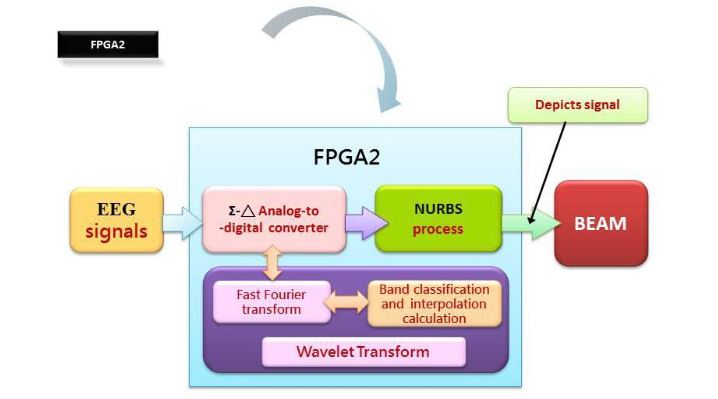
Figure 7. An embedded chip, FPGA2, design approach
Over recent years, the most popular digital signal processing approaches include Fast Fourier Transfer (FFT), Wavelet Transform (WT), Bispectral, Power Spectral Density (PSD), etc., among which adopted in this work are PSD and FFT, the most common approach seen in conventional brain wave analysis. A time domain signal is transformed into the frequency domain by means of FFT for synthesis of an EEG signal (see Figure 7).
CONSTRUCT THE BEAM

Figure 9. Sleep stage recognition according to brain wave
Saw tooth wave: As a feature of REM sleep, it is a wave with lower amplitude. Demonstrated in Figure 9 are the analysis results, provided by the Gerontechnolgoy Research Center, Yuan Ze University, Taoyuan, Taiwan, based on brain waves measured with P-Series PS2. Within the blue frame, the types of brain waves are indicated and the percentages there of at any time instant are recorded as well, while within the red frame, brain wave feature extractions are made, i.e., spindle, K-complex waves and REM, for the identification of stages 1, 2, NREM sleep and REM sleep.
TO USE MODIFIED ICA CAPTURE MORE PRECISE PHYSIOLOGICAL SIGNALS
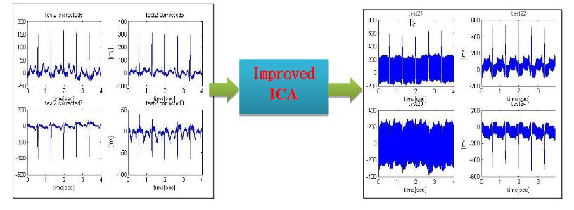
Figure 10. A superior physiological signal acquisition through an improved ICA algorithm
As suggested by Stogbauer et al. in 2004, ICA is modified to improve the quality of fetal electrocardiogram in an effort to separate fetal heartbeat from its mother’s. Since a conventional ICA algorithm might fail to recover the original signal, a modified version is employed in this work to reach the goa (see Figure 10).
THE FUZZY INFERENCE C4.5 CLASSIFICATION ALGORITHM TO CONSTRUCT BEAM SYSTEM DATABASE
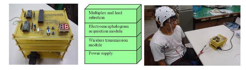
Figure 11. Photos for the presented EEG system and in a practical test
As expected, a SOC is built, compromising an integration of a real time EEG and BEAM diagnostics. Over conventional EEG approaches, it demonstrates the advantage of a high accuracy, a low error rate, an easy to use interface and a diagnostic expert database. This work is applicable to patients with epilepsy, encephalitis, brain tumors, cerebral, nervous headache, cerebral ischemia, cerebral hemorrhage, traumatic brain injury, neurasthenia, and so on (see Figure 11).
RESEARCH RESULTS AND EXPERIMENTAL ANALYSIS
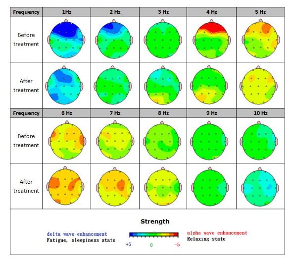
Figure 12. BEAM comparison between before and after treatment
The two brain maps shown in Figure 12 reflect patterns of an eleven-year-old developmentally delayed boy. Before treatment, a highly limited verbal ability was seen with no spontaneous speech. Along with significant attention problems, his visual perceptual skills suffered as well. As the treatment is continued, a noticeable stride has been made in several areas.
CONCLUSIONS AND FUTURE WORKS
BrainIC is developed as an auxiliary diagnostic tool to improve the accuracy of EEG analysis. The information contained in an EEG is displayed in colors in this work as a consequence of the integration of chip design with signal processing into neurological science field. As a rule, a quantitative analysis is made through BEAM, other than EEG, for accurate diagnosis of complicated diseases.
In an attempt to speed up a diagnostic process and improve the accuracy there of, a maximum number of electrode pads are placed in compliance with an international standard, a high speed and high precision Σ-Δ AD converter is adopted, a spectral analysis of an EEG signal is made by taking FFT, bands are classified and interpolated, and BEAM is plotted in the end. This study enables medical staff to acquire EEG signals in a more accurate manner, and the analysis thereof is made more efficiently as well.
Taking evoked potential analysis as an instance particular in the field of clinical anesthesia, evoked potential monitoring by BrainIC serves as an objective measure of the inhalation anesthetics central inhibition. Additionally, one would be diagnosed as a ddiction, epilepsy, attention deficit hyperactivity disorder, stroke, Alzheimer’s disease, multi-infarct dementia, and so forth by quantitative BEAM analysis.
In an effort to build a real time, portable multi-purpose system, an FPGA based PXA270 embedded platform is adopted as a physiologic signal measurement system, while a KL-720 system is employed as a front end, and real time data aggregation, classification, learning and validation can be made through a linkage to the embedded system via network. The greatest challenge in this work is to develop an FPGA based real time physiological signal acquisition system that involves a Σ-Δ AD converter, a differential amplifier, an FIR filter, and so on.
Subsequently, feature extractions are made from received physiological signals through an ICA algorithm. A complete visual platform, all the way from a prototype design to an embedded system, is developed with an NI LABVIEW interface. The signal processing involves an FFT, band classification, interpolation and NURBS to plot a BEAM. In the end, a diagnostic database is accurately built for analysis and inquiry purposes through all the existing BEAMs describing various types of diseases.
Source: National Chin-Yi University
Authors: Wen-Tsai Sung | Jui-Ho Chen | Kung-Wei Chang